
Research
The Bi Lab studies the biology of meningiomas, pituitary tumors, gliomas, brain metastases, and other skull base tumors, combining genomics, imaging, and outcomes research to sharpen diagnosis, guide treatment, and make surgery safer. Below are a few of our active projects, organized by disease, and the research directions each one draws on.
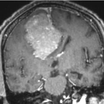
Meningioma
Meningiomas are the most common primary brain tumor, and most are slow-growing and curable with surgery. But the standard grading system, based on how a tumor looks under the microscope, doesn't always predict how an individual tumor will behave. We are working to build a more precise molecular fingerprint for each meningioma, so that a patient's risk is defined by their tumor's biology, not just its appearance.
In a study of more than 1,000 meningiomas published in Nature Communications, Ruchit Patel and colleagues mapped how tumors gain or lose pieces of chromosomes, and showed that small differences in how these changes are measured can shift a tumor between a low- and high-risk category for as many as 1 in 5 patients. This work is helping refine the thresholds pathologists use to grade tumors.
We also asked whether Ki-67, the standard marker doctors use to gauge how fast a tumor is growing, was measuring what everyone assumed. Using single-cell technology across more than 120,000 individual cells, Xiaopeng Guo and colleagues found that Ki-67 is produced not only by tumor cells but also by immune cells within the tumor, especially in lower-grade disease, and that this balance shifts with tumor grade, prior radiation, and patient age. That means the same Ki-67 result can mean different things from one tumor to the next.
Most recently, work led with Zach Moynihan showed that a meningioma's immune environment is not fixed: it evolves as the tumor's biology changes, including after treatment. Andrew Dunbar, a medical student in the lab, is now helping extend this work, combining genomic and immune fingerprinting to understand how treatment can reshape a tumor's biology, with the goal of identifying which tumors are truly high-risk and which patients might safely need less treatment.
- Distribution of copy number alterations and impact of chromosome arm call thresholds for meningioma · Nature Communications
- Ki-67 in meningioma: distribution and implications · Journal of Neurosurgery
- Meningioma microenvironment harbors a rich immune landscape that evolves with biological state · Neuro-Oncology
Research team: Ruchit Patel, Xiaopeng Guo, Zach Moynihan, Andrew Dunbar.
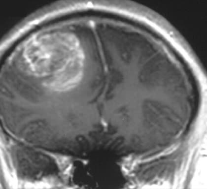
Glioma
Gliomas are the most common cancerous brain tumor in adults, and how a glioma is classified and treated now depends on genetics as well as how it looks under the microscope. To make sure these updated classifications reflect real-world outcomes, Hia Ghosh and Ruchit Patel led an analysis of 4,400 glioma patients, published in Neuro-Oncology. They found that survival for most glioma subtypes is meaningfully better today than older reference studies suggested, and built updated, clinically applicable tools that combine a tumor's molecular profile with a patient's clinical picture and treatment to give a more accurate, individualized prognosis.
A related study looked specifically at IDH-mutant astrocytomas, a common glioma subtype, to see whether genetics could sharpen risk beyond grade alone. Analyzing nearly 1,000 patients, the team found that loss of a gene called CDKN2A/B, along with certain chromosomal amplifications, identified patients at substantially higher risk regardless of how the tumor appeared under the microscope: patients with an intact CDKN2A/B gene and no amplifications lived, on average, more than 17 years after diagnosis, compared with under 3 years for those with CDKN2A/B loss in a grade 4 tumor. These findings are helping refine how we counsel patients and plan treatment.
As these molecular tools evolve, so does surgery itself. The lab's intraoperative neuromonitoring (IONM) program tracks the function of critical nerve and brain pathways in real time during glioma resection, helping the surgical team remove as much tumor as safely possible.
Research team: Hia Ghosh, Ruchit Patel.
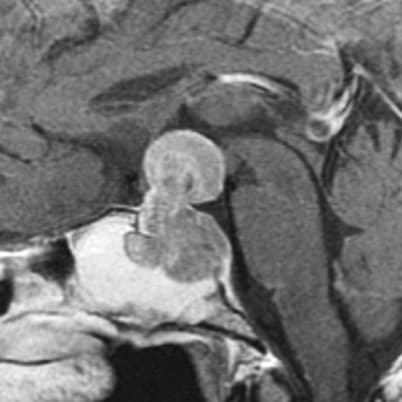
Pituitary Tumors
Most pituitary tumors are benign, but a subset behave aggressively and resist standard treatment. Over the past decade, the lab has mapped the genomic drivers of these tumors, from early work identifying recurrent oncogenic mutations and copy-number changes in pituitary adenomas, to more recent profiling of how immune cells interact with pituitary tumor cells and express immune checkpoint molecules.
Building on that foundation, the team is now characterizing the pituitary tumor microenvironment in greater detail: the non-tumor cells, blood vessels, and signaling molecules that surround and interact with tumor cells, to understand what allows some pituitary tumors to grow aggressively or resist treatment, and to identify new therapeutic targets. This work is ongoing and not yet published.
Research team: Zach Moynihan, Sydney Wiredu.
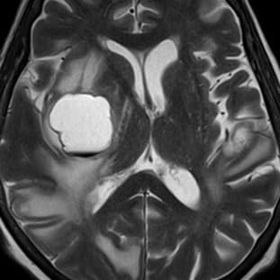
Brain Metastases
Where a patient with brain metastases is treated, and how well-connected that hospital is within the broader network of cancer care, can meaningfully affect outcomes. In a study published in JAMA Network Open, Lilin Tong and colleagues used physician-referral network analysis to show that hospital “connectedness” is associated with outcomes for patients with brain metastases. Related work from the lab has mapped how patients are transferred between hospitals for brain metastases and other acute neurosurgical conditions across Massachusetts.
Andrew Dunbar, Lilin Tong, and Justin Kim are extending this line of work, applying network analysis to understand how the structure of a patient's care network shapes brain metastasis outcomes, with the goal of identifying points in the system where better connectivity could improve care.
Zsombor Gal is also leading a related study on salvage surgery: a second operation considered when a brain metastasis regrows after initial treatment. Analyzing 207 patients who underwent salvage surgery for 234 recurrent brain metastases, the lab's largest study of its kind, he found that a complete resection, achieved in about two-thirds of cases, substantially lowered rates of tumor regrowth, need for further treatment, and death, and improved patients' odds of symptom relief and coming off steroids. A patient's overall health going into surgery mattered just as much, across nearly every outcome measured.
- Role of hospital connectedness in brain metastasis outcomes · JAMA Network Open
- Interhospital transfer dynamics for patients with intracranial hemorrhage in Massachusetts · Frontiers in Neurology
Research team: Andrew Dunbar, Lilin Tong, Justin Kim, Zsombor Gal.
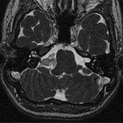
Epidermoid Cysts
Epidermoid cysts are rare, slow-growing tumors thought to arise from skin cells trapped during early development. Most are benign, but in very rare cases they can become locally aggressive or transform into cancer, and almost nothing is known about what drives that process.
Gabrielle Luiselli and Ellie Shahbo are developing an animal model of epidermoid cyst formation alongside molecular profiling of patient tumor samples, aiming to identify the genetic changes that drive cyst growth and, in rare cases, malignant transformation. This work is currently being prepared for publication.
Research team: Gabrielle Luiselli, Ellie Shahbo.
Imaging
Across all of these tumor types, safe surgery is predicated on detailed knowledge of the relevant anatomy. We apply advanced imaging modalities to assess critical structures that may be encountered during the operative approach, to improve preservation of neurovascular function.